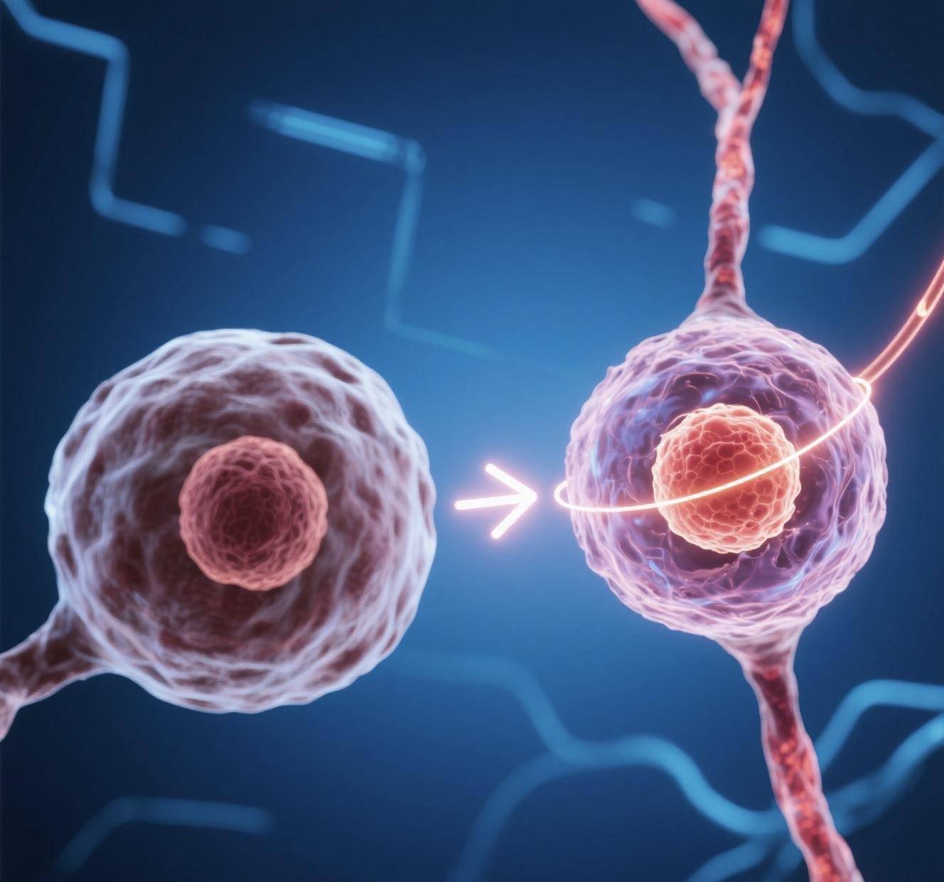
In a landmark study published in the new issue of nature, the Stanford University School of Medicine team used healthy precursor cells that were not genetically matched, replacing more than half of the microglia in mice with Sandhoff disease extended the animals’ life span from 135 days to 250 days, and their motor function and exploratory behavior returned to almost normal levels. It is also the first to provide a“Ready-to-use” blueprint for cell therapy for currently incurable fatal brain diseases, such as Tessak’s disease and Sandhoff’s disease.
Tsax and Sanderhoff are lysosomal storage disorders. Due to a lack of key enzymes and a build-up of metabolic waste in the nerve “Scavenger” microglia and adjacent neurons, the child degenerates rapidly within months of birth and usually dies before the age of two. Previous attempts of Hematopoietic stem cell transplantation required systemic chemotherapy to clear the marrow, and healthy cells were difficult to cross the blood-brain barrier, with a success rate of less than 30% and associated with rejection or graft-versus-host reaction.
The team used a “Brain-specific transplantation” strategy, which involved exposing the brains of mice to low doses of radiation followed by drugs to temporarily remove the microglia, microglial precursor cells from unmatched donors were then injected directly into the ventricles, followed by two approved immunomodulatory drugs to block peripheral immune attacks. The results showed that the new cells still accounted for more than 85% of the total microglia in the brain after eight months and did not spread to other parts of the body.
The behavioral tests were equally exciting: all of the untreated mice died within 135 days, while the five mice that received the transplants were still alive at the end of the experiment; not only did they dare to enter the center of the open field, the grip strength of hind limbs was also significantly better than that of the control group. Histological analysis revealed that lysosomal enzymes secreted by donor microglia were taken up by neighboring neurons, suggesting that the “Cell-out” mechanism may be key to therapeutic efficacy.
The results also solved three problems: no systemic toxicity pretreatment, no gene editing to supplement the deleted enzyme, and to avoid rejection. The radiation dose, microglia scavenger and immunosuppressant used in the protocol have been used in other diseases and have the potential of rapidly entering clinical practice. At the same time, the therapy does not rely on the patient’s own cells, and is expected to become a“Shelf product” like blood transfusion in the future, greatly reducing the cost and waiting time.
The team points out that common Neurodegeneration such as Alzheimer’s and Parkinson’s are also associated with microglial dysfunction, which may be a “Slow version” of lysosomal disease. If follow-up human trials are successful, the beneficiaries will be millions of Neurodegeneration, not just children with rare diseases. Next, the team plans to validate the safety of this therapy in larger animal models that are closer to humans, and to discuss the design of early clinical trials with the U.S. Food and Drug Administration.
Editor-in-chief points
Cell therapy attempts to replace damaged neurons with healthy cells or activate endogenous repair mechanisms, offering new hope for the treatment of lysosomal storage disorders and Neurodegeneration. In the latter, for example, Neurodegeneration such as Alzheimer’s and ALS are still in desperate need of more effective drugs and treatments. The new research adopts the strategy of“Brain region-specific transplantation”, which not only avoids systemic toxicity pretreatment, but also avoids rejection. It provides a new way to treat this kind of disease by cell therapy. In the future, cell therapy, combined with cutting-edge technologies such as gene editing and targeted delivery, is expected to have greater potential in related fields of medicine.